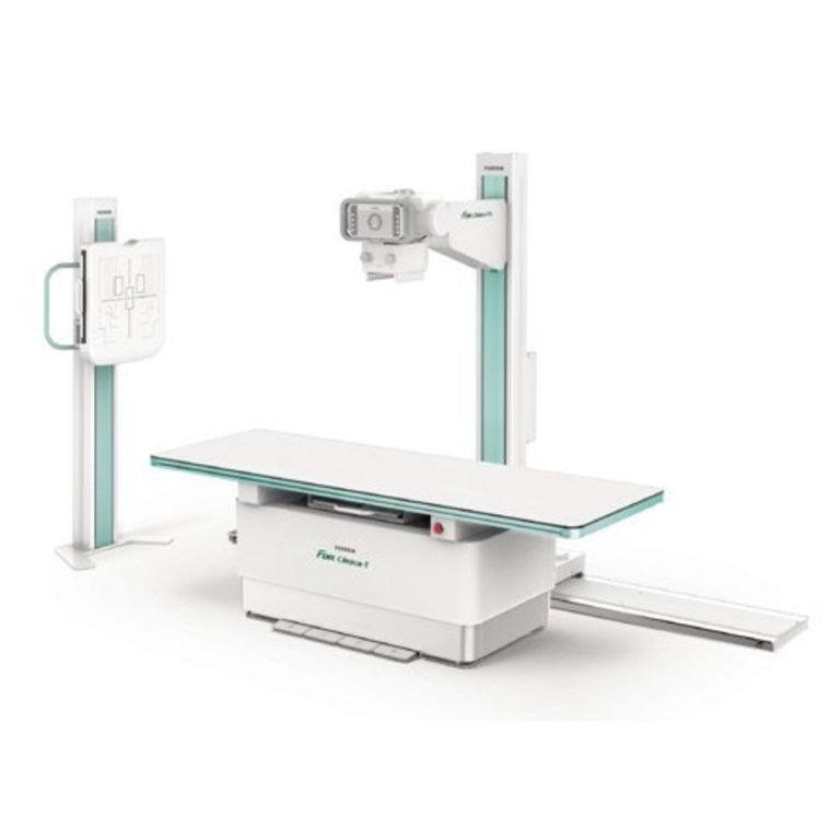
The cutting edge headway has likewise impacted radiology. A long time back, radiology was restricted to X-Rays. However, throughout the long term, traditional systems have seen a huge improvement, prompting upgraded insight of treatment for a great many individuals across the globe. Of the latest accomplishments, the conspicuous ones can be referenced as — Digital Radiography and Digital Mammography, Ultrasonography, Remote review framework, CT Angiography, Replacement of Exploratory Surgery, and so forth.
Advanced Radiography and Digital Mammography
Advanced radiography has immensely further developed the X-Ray quality, and speed and supplanted all the exhausting darkroom strategies, as these can be shared promptly for fast indicative outcomes. The regular X-Ray used to take about 30 minutes for the entire method while the computerized radiograph can be obtained with only a tick of a button while conveying far superior quality.
USG: Ultrasonography chips away at the standard of getting reverberation from reflected sound waves. It has progressed massively throughout the years from 2D to 4D pictures and remote review frameworks of the current day. USG is a vital imaging methodology. It’s unique in relation to other imaging methodology as it is continuous and more secure, as it doesn’t include radiations. USG has additionally progressed over the course of the years with dopplers, directed needle situation elastography and some more.
Remote survey framework
Radiology has progressed enormously throughout the years yet the miserable truth is that in India and numerous different nations many individuals don’t approach fundamental radiological techniques.
Kenya with a populace of 43 million has just 200 radiologists. In rustic Nepal, there are spots where individuals travel very nearly two days for getting an X-Ray. Online frameworks permit doctors to send and get pictures and reports from everywhere in the world.
CT Angiography
Conventional angiography requires a few hours, requires tranquillizers and could in fact make harmful vessels. Be that as it may, CT-Angiography requires 10-15 minutes without such dangers. It tends to be utilized to picture any conduit in the body including the cerebrum, coronary, lung, kidney and so on with late advances in CT angiography. It gives an image which is three-layered and resembles a genuine blood vessel framework in our body and is well disposed of so that clinicians might see the pathology.
Substitution of Exploratory medical procedure
CT examination lessens negative appendectomy. It is vital in injury patients where a choice must be taken from CT regardless of the patient’s necessities medical procedure or not. So lessening injury brought about by a medical procedure and saving a ton of patients’ use performed on superfluous medical procedures.
X-ray
X-ray assumes a pivotal part in recognizing pathology at its earliest. In stroke patients, it can identify the progressions found in Acute infarcts (a little restricted area of dead tissue coming about because of disappointment of blood supply) at its earliest.
Late advances in MRI like spectroscopy magnetisation move and utilitarian MRI assume a significant part in choosing the administration and kind of treatment. X-ray is radiationless and gives exceptionally exact and minute subtleties of pathology.
PET – CT
Positron emanation tomography went on with CT check pictures gives simple discovery of even little growths and metastatic data and furthermore give the metabolic status of cancer as doctors get a superior thought of what is happening and how to appropriately treat it and furthermore help in observing chemotherapy.
Competence in radiography
In the field of radiologic innovation, one part of the calling requires able abilities in radiographic openness factor procedure. The said capability is fundamental, particularly in the demonstrative X-Ray imaging, wherein openness factors are the way to exact conclusion and giving radiation measurement to least even out. For a long time, film-screen strategy has been the technique for decision in radiographic imaging (Bushong 2009). The film-screen framework utilizes radiographic movies, radiographic escalating screens and wet science to make the picture appears. Besides, this customary framework ought to stick to the principles of the darkroom prerequisites. The film-screen framework has a similar idea as a common traditional camera. In a film-screen method, radiologic technologists ought to be sure on the openness variables to be applied in a specific openness on the grounds that ill-advised determination of openness elements can prompt overexposure or underexposure of the film. Overexposure or underexposure corrupts picture quality and hence, it can prompt dismissal of film, subsequently requiring the requirement for rehash assessment. Rehash assessment gives a pointless portion to the patient and extra expenses for the division.
Then again, similarly to different advancements in innovation, demonstrative imaging has changed its direction from customary to computerized. PC applications are utilized these days in demonstrative imaging modalities. A proper similarity that is simple for the vast majority to comprehend is the supplanting of commonplace film cameras with computerized cameras: pictures can be taken, promptly inspected, erased, revised, and trimmed, and in this way shipped off an organization of PCs. A registered radiography framework (CR) is a reasonable answer for advanced imaging. Rather than the film, CR utilizes an imaging plate to catch X-Rays and makes it noticeable when the plate is filtered into a PC and digitized it. When the picture is switched over completely to information, it very well may be recorded on a laser-printed film or can be sent and put away carefully. It has extraordinary highlights like control or upgrade of the picture. Its particular programming is utilized to picture seeing with improved capacities like film-screen framework, like differentiation, splendour, and zoom. (dicomsolutions.com, 2011).
Registered radiography enjoys pragmatic specialized benefits contrasted and customary methods, for example, wide difference dynamic reach, post-handling usefulness, different picture seeing choices, and electronic exchange and filing prospects. In this framework, picture quality can be accomplished in light of the post-handling methods that are impractical with the film-screen framework. This framework is advantageous for the technologists in light of the fact that the RT can make up for openness strategy mistakes by changing the procedure during the post-handling period of the picture as opposed to that season of openness.
Also read- www.strategicmarketresearch.com
